This is a HIPAA de-identified open-online-patient-record with patient information posted here after collecting informed patient consent.
History presented by PGY1 Dr Su,
65 yr old man came with complaints of abdominal distention associated with respiratory discomfort and pedal oedema since 20 days , gradually progressive and also complaints of loss of appetite and constipation since 15 days.
Vitals : PR 105bpm,BP 140/100,RR 22cycles/min,spO2 94%,
On examination abdomen distended with umbilicus everted,fluid thrill present,on auscultation basal crepts present.


Ascitic fluid aspirate

Cell count of ascitic fluid aspirate showing 5000 neutrophils per hpf
 Ascitic fluid aspirate growing E coli


History presented by PGY1 Dr Su,
65 yr old man came with complaints of abdominal distention associated with respiratory discomfort and pedal oedema since 20 days , gradually progressive and also complaints of loss of appetite and constipation since 15 days.
Vitals : PR 105bpm,BP 140/100,RR 22cycles/min,spO2 94%,
On examination abdomen distended with umbilicus everted,fluid thrill present,on auscultation basal crepts present.


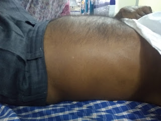
Distended abdomen

Ascitic fluid aspirate

Cell count of ascitic fluid aspirate showing 5000 neutrophils per hpf


Therapeutic aspiration

CECT abdomen
Ascitic fluid aspirate culture sensitivity
Conversational decision support system
Web Log Medicine:
[1/28, 7:34 PM] Rb:
https://jamanetwork.com/journals/jamainternalmedicine/article-abstract/607185
"In none of four episodes of secondary bacterial peritonitis was there an exponential decline in neutrophil count after antimicrobial therapy was initiated. In fact, the first follow-up neutrophil count was greater than the baseline value in all four episodes. The response pattern of the ascitic fluid neutrophil count to antimicrobial therapy is helpful in differentiating spontaneous from secondary bacterial peritonitis."
[1/28, 7:44 PM] Rb: Does our patient's ascitic fluid satisfy Runyon's criteria?
"Runyon’s Criteria for secondary bacterial peritonitis requires two of these three features:
total protein >1 g/dL, glucose <50 mg/dL (2.8 mM), and lactate dehydrodgenase above the upper limit of normal for serum."
http://emcrit.org/pulmcrit/secondary-bacterial-peritonitis/
[1/28, 7:52 PM] Rb: This 👆article is quite informative.
[1/28, 9:24 PM] VL AP: Good evening Sir, even abdominal tuberculosis needs to be ruled out...
Irrespective of raised neutrophils counts...
[1/28, 9:29 PM] Rb: How do we rule it out short of a peritoneal biopsy? The ADA may help to rule it in slightly although may not rule it out. Again with the predominant neutrophils in the ascitic fluid it's unlikely that ADA will be high
[1/28, 9:32 PM] VL AP: Sir if no ADA can we go with large volume ascitic fluid culture and montoux....
[1/28, 9:39 PM] Rb: Culture ascitic fluid for mtb? Yes Mantoux can be done but will only tell us about infection but not whether he is diseased
[1/28, 9:39 PM] VL AP: Ok sir...
[1/28, 9:41 PM] VL AP: Sir how about malignancy as one of the differential as he is elderly..
Ascitic fluid for malignant cell ...
[1/28, 9:49 PM] Rb: Yes cytology was done as shown in above slide image. No malignant cells were seen. The Usg doesn't show any obvious malignancy. CT abdomen is awaited
[1/28, 9:49 PM] VL AP: Ok sir
[1/28, 10:35 PM] PGY 1 AS: https://www.ncbi.nlm.nih.gov/pmc/articles/PMC5348596/
[1/28, 10:35 PM] PGY 1 AS: This was a case of a secondary peritonitis without perforation that was treated successfully with antibiotics and surgical intervention. Although the case did have certain characteristics of SBP, such as monomicrobial infection and improvement of absolute neutrophil count after 48 h of antibiotics, persistence of patient symptomatology prompted further evaluation to look for an alternate diagnosis. Runyon’s criteria have an estimated sensitivity and specificity for predicting secondary bacterial peritonitis of 67 and 96%, respectively and our patient met the criteria. That together with the lack of improvement prompted us to probe further. A repeat paracentesis is not necessary for all patients with infected ascites but should be considered in patients with one or more characteristics of secondary peritonitis as detailed above. Re-categorizing this case from SBP to secondary peritonitis allowed us to advocate for a likely curative surgical intervention.
[1/29, 6:21 AM] Rb: Thanks Aditya. I guess you quoted this from a similar case as hours. Based on this and other case based evidence what do you propose should be done for our current case?
[1/29, 11:10 AM] PGY 1 AS: It looks like it's not satisfying Runyon's criteria, so there's a 98% chance it's secondary peritonitis.
Web log central:
[1/30, 9:28 AM] Microbiologist: E.coli isolated in peritoneal fluid. Sensitive to gentamicin, amikacin & Meropenem.
[1/30, 11:58 AM] MS: So what is the clinical status of this patient with bacterial peritonitis at present.
[1/30, 12:48 PM] Rb: Subjectively he's been doing well on antibiotics although not completely out of the woods. He still has mild features of peritonism and we are currently thinking it is perhaps spontaneous bacterial peritonitis rather than secondary bacterial peritonitis. He's on cefotaxime. Ma'am can you share the deidentified complete report of his sensitivity? Can his CECT films be shared and web logged by the radiology department here? We are also trying to repeat his ascitic tap to see if there is any reduction of the WBC counts after 48 hours
Web log Medicine
[2/1, 8:36 AM] Rb: Answering my own question to the surgeons
Therapeutic large volume ascitic fluid drainage (aka "large volume paracentesis) is not an absolute contraindication and can be a tolerable and safe therapy in some selected cirrhotic patients with tense ascites and SBP."
https://www.ncbi.nlm.nih.gov/m/pubmed/12499817/
[2/1, 8:36 AM] Rb: Prof K as mentioned here, can you further elaborate if the counts would be erroneously high or erroneously low because of the clot.
The patient appears slightly clinically better with antibiotics although not completely out of the woods. The CECT abdomen is normal except for large ascites. My question to surgeons here is what would be the role of therapeutic ascitic drainage in such a patient where there is a persistent dilemma between spontaneous and secondary bacterial peritonitis? Dr Sufiya can you elaborate on his alcoholic liver disease with multinodular cirrhosis diagnosis in terms of history and clinical findings?
[2/2, 1:21 PM] +91 90308 17067: We aspirated 600ml fluid sir
[2/2, 1:22 PM] Rb: Good. 👍 Your name is not visible here
[2/2, 1:25 PM] +91 90308 17067: Am ur unit Intern sir 🙂
[2/2, 1:38 PM] Rb: 👍
[2/3, 7:44 PM] Rb: Yes do you remember the discussion we had around this peculiar phenomenon in sepsis of platelets going up even as the WBC counts were going down? Do you remember which paper the Pathology PGs had shared that day? Can we revise it?
[2/3, 7:46 PM] Rb: It was around the last sepsis patient we had presented in the morbidity meeting (by Dr SL).
[2/3, 8:26 PM] Rb: Dr S I had a discussion with Dr M, and he felt we can try to check out for perforation by introducing methylene blue dye through his Ryle's tube and checking if the ascites fluid turns blue. Vivek any articles around this you can find and share?
[2/3, 10:13 PM] Vivek Poddar 3: Oh sorry. Saw it now.
[2/3, 10:16 PM] Vivek Poddar 3: "Dye studies may be useful for evaluating patients with a pleural effusion and a thoracostomy tube who are suspected to have an esophageal leak. Methylene blue introduced cautiously via a nasoesophageal tube will make or confirm the diagnosis by causing blue discoloration of the chest tube drainage." UTD
[2/3, 10:16 PM] Rb: None for intestinal perforation?
[2/3, 10:19 PM] PGY3 SR: https://www.ncbi.nlm.nih.gov/pubmed/20038507
Full strength 1% MB dye instilled into the gastric lumen can potentially be used as a marker for detection of mucosal perforations of 1.2 mm or greater during laparoscopic pyloromyotomy.
[2/3, 10:23 PM] Vivek Poddar 3: "Methylene blue can reliably be used as routine diagnostic test for the assessment of upper gastrointestinal integrity, where bacterial load is low. In the lower gastrointestinal tract, where bacterial load is generally higher, the dye can indicate leaks, only if extended intestinal passage after oral ingestion is avoided. In all other cases, the examiner has to be aware of false-negative results by bacterial discoloration of methylene blue."
https://www.ncbi.nlm.nih.gov/m/pubmed/16260876/
[2/3, 10:25 PM] Rakesh Biswas: 👍
[2/3, 10:27 PM] Vivek Poddar 3: https://bmcsurg.biomedcentral.com/articles/10.1186/1471-2482-7-15
I am not understanding if integrity of colonic anastomosis has any relation with perforation!?
[2/3, 10:30 PM] Rb: Dr S did we repeat his ascitic fluid culture in Saturday?
[2/3, 10:31 PM] S: Yes sir I sent the sample
Minutes of Face to face morbidity meeting 2/5
The dilemma was shared and discussed with medical college faculty from different departments accumulated in a mini auditorium that Tuesday and most surgeons were unanimous in voting against perforation and by our above E logged discussion we too knew that the therapeutic response to antibiotics was an important factor to suggest that what he had was likely to be spontaneous bacterial peritonitis.
A CECT abdomen was repeated with oral contrast and it didn't reveal any leakage into the peritoneum.